Astrobiology Revealed #20: Lara Maldanis
on uncovering putative Mars-like microfossils at Rio Tinto
by Aubrey Zerkle
For this Astrobiology Revealed, we asked Lara Maldanis about her paper “Unveiling Challenging Microbial Fossil Biosignatures from Rio Tinto with Micro-to-Nanoscale Chemical and Ultrastructural Imaging.” Lara is currently a Marie Curie Postdoctoral Fellow at the Earth Sciences department of the VU Amsterdam. Lara discusses how high-resolution 3D imaging methods like the ones they used could be the key to finding definitive traces of life in rocks from challenging environments like Mars. (This interview has been edited for length and clarity.)
How did you become interested in astrobiology in general, and the search for Martian microfossils in particular?
My journey into astrobiology started with paleontology during my master’s, when I was searching for fossilized hearts in exceptionally well-preserved fossil fishes from Brazil. At the time, I got exposed to some very cool methods based on X-ray imaging using synchrotron accelerators, which could reveal impressive details of fossilized organisms. I decided to pursue a PhD at the intersection between these physical methods and some challenging questions in paleobiology. I was particularly triggered by the controversies regarding some of the oldest fossils on Earth, Archean microfossils.
The search for potential fossil microbes on Mars relies on our knowledge of how these structures can be preserved and especially on how we can identify them – a topic that’s still controversial when debating the oldest fossils of our planet. So for my PhD, I aimed to contribute to this challenge by exploring a new and very sensitive synchrotron-based X-ray nanotomography method being developed in other fields, such as physics and material sciences, and adapt it to studying very old microorganisms preserved in rocks. Since then, I have been passionate about exploring new technologies that can be used to study fossil biosignatures on Earth and which could, in the near future, be able to reveal potential traces of life preserved in samples returned from Mars.
Why did you decide to study the terraces at Rio Tinto?
Rio Tinto is a fascinating environment where a surprising diversity of extremophiles thrive despite its extremely acidic waters rich in toxic metals. Iron-rich deposits in this setting contain some iron-sulfate minerals such as jarosite— a mineral that the Opportunity rover discovered in the terraces of Meridiani Planum on Mars, where similar iron-rich and acidic terrains existed. These Martian deposits date back approximately 3.5 billion years, around the same age as Earth's oldest known fossils. The presence of both living and fossil microbes in Rio Tinto provides a valuable opportunity to study how life endures—and how it might be preserved as fossils—in the kind of acidic and iron-rich environments that once existed on Mars.
In the article you mentioned that Rio Tinto has "chemical conditions commonly thought to be challenging for life and fossil preservation," but you found an impressive set of microbial structures nonetheless. Could you briefly describe these challenging conditions, and how these microfossils overcame them?
Hematite-rich terrains are considered thermodynamically unfavorable for the preservation of organic matter, which is one reason this type of environment was initially deprioritized in the search for fossils on Mars. However, a diverse assemblage of fossils has been found preserved in rocks from Rio Tinto, including fossil microorganisms, which challenged this paradigm. Using highly sensitive 3D imaging techniques at nanometer resolution, we revealed that these fossils consisted of hollow structures with well-preserved, consistent 3D filamentous shapes, yet they contained no traces of cell envelopes or organic remains. Through additional analyses, we concluded that they were preserved as 3D mineral casts or microborings. While the absence of organic material might seem to argue against their biological origin, we identified multiple structural and contextual features that strongly supported their authenticity as fossils.
You examined these microfossils at multiple scales using several types of synchrotron and X-ray imaging techniques. What are the main advantages of your methods versus traditional methods for examining putative microfossils?
Conventionally, microfossils have been studied using optical microscopy and, in recent decades, with more advanced imaging techniques such as electron microscopy, Raman spectroscopy, and confocal laser scanning microscopy, among others. However, visible-light-based methods lack the resolution needed to reveal details of fossilized bacteria, which can be smaller than 1 micrometer in diameter. This makes it difficult to observe cellular morphology in detail or to distinguish these structures from other similar ones in rocks.
Electron microscopy can overcome this resolution limit, but electrons do not penetrate deeply into the rock, revealing only fossil fragments at the surface or in very thin (∼100 nm) layers. Achieving 3D imaging with electrons became possible through focused ion beam scanning electron microscopy (FIB-SEM), but this technique is destructive, costly, and therefore limited in both the study area and the spacing between consecutive images.
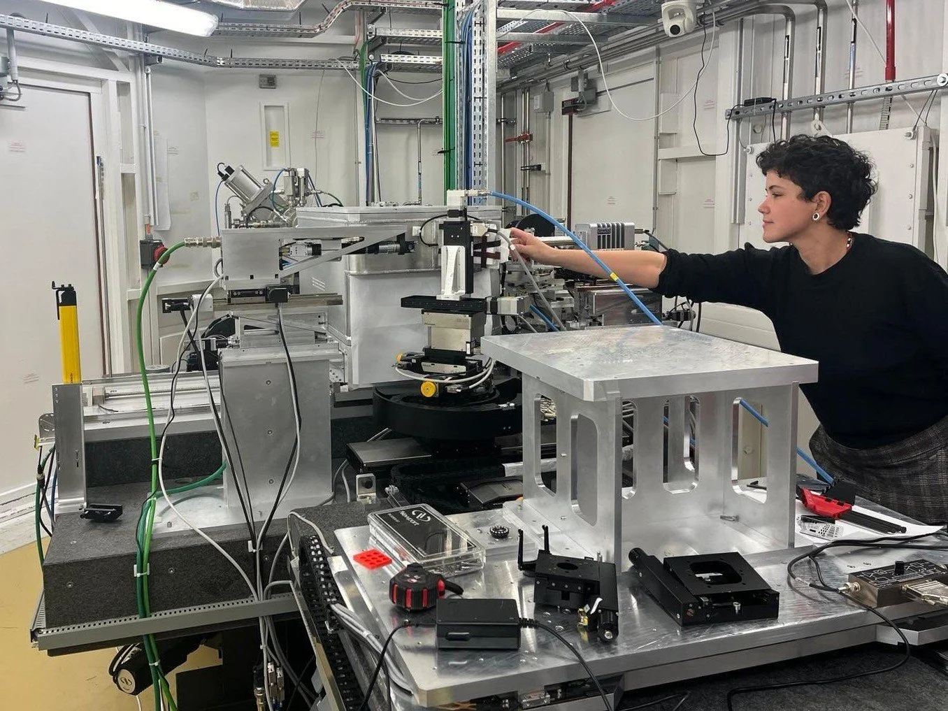
X-ray nanotomography offers a non-destructive approach for imaging microfossils in 3D, overcoming these limitations. The X-ray method I explored in this work, Ptychographic X-ray Computed Laminography, is based on the technique I used in my PhD but has been developed to analyze samples that cannot be rotated 360°—a key requirement for standard tomography that restricts sample size and shape. This innovation enables high-resolution 3D imaging of large specimens, such as the rock thin sections used for classical and preliminary optical microscopy investigations. Since the sample extends laterally, different regions can be explored and imaged at multiple scales, ranging from a few micrometers to hundreds of micrometers. This enables nanometric-resolution investigations of multiple specimens, their interactions with one another, and their relationships with surrounding minerals, providing valuable insights into their metabolism, ecology, taxonomic affinity, and fossilization processes.
We also explored complementary chemical information using synchrotron nano X-ray fluorescence, a highly sensitive method capable of detecting ultra-trace levels of metals. Despite this sensitivity, no elements were found associated with the fossils, supporting their cast nature.
What type of microbe-mineral interactions did you find, and how relevant are they for putative Martian life?
One important thing about microbes is that they are never alone—they always exist in communities, constantly interacting with other species and affecting their surroundings. This means that if we find a possible fossilized microbe in a Martian rock, examining its context is just as important as studying the microbe itself.
For example, in our study, we observed that in addition to the larger microbial filaments visible under optical microscopy, there were also smaller specimens—too tiny to detect by optical microscopy—closely interacting as part of a multispecies assemblage. Some microbial filaments were cross-cutting a dense cluster of mineral particles. These filaments were exceptionally well-preserved in 3D, indicating that they could not have been passively transported into the mineral pack during the sediment remobilization —otherwise, their delicate morphology would have been disrupted. Instead, this suggests that these microbes arrived later in the diagenetic process, actively “diving” into the crystal pack—likely in search of nutrients.
By reconstructing interactions like these, we gained insights into their ecology, metabolism, and fossilization processes. This contextual evidence was particularly crucial since, as mentioned earlier, these fossils lacked any preserved cell structures or organic material. Without this context, they could hardly be recognized as microfossils—something that may also apply to structures preserved in other harsh, challenging environments, such as those on Mars.
Based on your results, what good news can you offer for astrobiologists looking for life on Mars?
The good news is that we can use new synchrotron methods to extract an unprecedented level of information from fossil microbes—even from extreme environments like Rio Tinto (and, hopefully, Mars!) These methods can overcome challenges such as detecting tiny specimens, identifying fossils without preserved organic material, and analyzing structures embedded in dense rocks or hidden within complex mineral networks. These advancements could be key in finally identifying definitive traces of life on Mars.
And what's the bad news?
The bad news is that this kind of analysis requires very large particle accelerators, and like many other highly sensitive techniques, it can only be performed on Earth. We will have to rely on sample return missions to bring rocks from Mars, hoping that the selected cores have correctly collected these traces. Additionally, these samples will be extremely precious, and the imaging methods that reach the nanoscale will always require some degree of sample preparation, which could present additional challenges. Not to mention the potential for radiation damage, which we will need to be especially careful about, particularly if organics are present!
Is there anything else you’d like to discuss that I haven’t asked you about?
We actually started this project looking for patterns of preserved metallome elements, inspired by your work from 2005! We were quite puzzled when we found no metals associated with the fossils and had to dig deeper to understand why. It was a fascinating process because fossils from different localities can vary so much depending on their geological history. Facing these challenges pushes us to be creative and explore new ways to investigate them. I think this kind of problem-solving is great preparation for future scientists analyzing samples brought back from Mars!


